Tissue oxygen (pO2) measurement by photoacoustic imaging
In this project we focus on developing the scientific and technological foundations for non-invasive imaging of oxygen distribution in cancer tumors. Low oxygen level (hypoxia) in tumors greatly inhibits cancer therapy and indicates a poor prognosis. Successful implementation of the technology would provide the oncologist with essential information to significantly improve the efficacy of both radiation therapy and chemotherapy. It would also lead to better understanding of cancer therapy mechanisms and assist in developing new hypoxia-targeted cancer therapies.
The underpinning technology that drives the project development is photoacoustic lifetime imaging (PALI). PALI is a pump-probe approach to photoacoustic imaging that extends it to allow measurement of transient absorption processes.
In order to apply PALI for tissue oxygen imaging, an oxygen-sensitive dye is used to stain the tissue. We use Methylene Blue which is a non-toxic dye, commonly used in other clinical imaging applications.
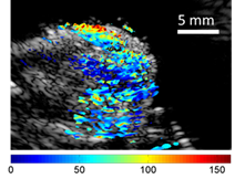
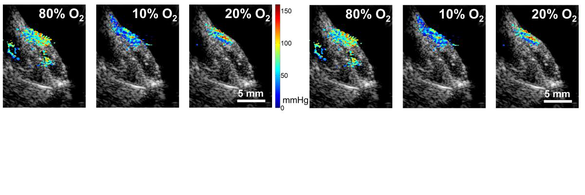
Our system combines PALI image with conventional ultrasound imaging, thus providing an anatomical reference frame for tissue-oxygen information. Sample images of a phantom object consisting of two tubings and a hind limb of a mouse bearing a cancer tumor (originated from a human prostate cancer cell line) are shown below.

In-vivo imaging: Dual-modality images of a hind limb of nude mouse bearing a tumor. Left: Photoacoustic image in red overlaid on ultrasound b-mode image. Right: PALI Tissue Oxygen image in color scale (spanning pO2 range of 0 to 150 mmHg) overlaid on ultrasound B-mode image. Ultrasound image is shown in gray levels spanning a range of 50 dB.
